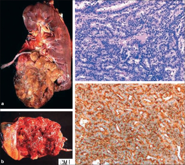
Figure 1.

(a) Renal carcinoid tumor. Grossly it forms a circumscribed mass with a yellow cut surface with foci of hemorrhage. (b) Occasionally, it can have a hemorrhagic and friable cut surface. (c) Microscopically it comprises ribbon and trabeculae of uniform tumor cells, (d) which are positive for synaptophysin immunostain
