
Figure 2. BDNF and NT-3 Stimulate Phosphorylation of Tyrosine Residues in the trkB Tyrosine Kinase Receptor gpl45trkB.
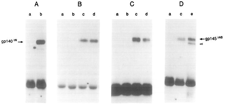
(A) gp140trk-expressing E25-48 cells incubated in the absence (lane a) or presence (lane b) of 50 ng/ml NGF.
(B) gp145trkB-expressing Z52-17 cells incubated with a 1:10 dilution of supernatant from mock-transfected COS cells (lane a), 50 ng/ml NGF (lane b), a 1:10 dilution of supernatant from COS cells transfected with pVN1 (BDNF) (lane c), or a 1:10 dilution of supernatant derived from COS cells transfected with pVN2 (NT-3) (lane d).
(C) gp145trkB-expressing Z52-17 cells incubated with supernatant from Sf9 cells infected with wild-type baculovirus (lane a) or 50 ng/ml NGF (lane b), 50 ng/ml baculovirus-derived BDNF (lane c), or 50 ng/ml baculovirus-derived NT-3 (lane d).
(D) Lane a, Z52-17 cells incubated with supernatant from Sf9 cells infected with wild-type baculovirus. Lane c, Z52-17 cells incubated with 50 ng/ml baculovirus-derived BDNF. Lane e, Z82-41 cells transformed by cotransfection of pFRK44 (trkB) and pLL42 (BDNF) DNAs in the absence of ligand.
All incubations were performed for 10 min at 37°C. Cells were lysed and incubated with rabbit polyclonal antibodies elicited against gp140trk (Martin-Zanca et al., 1989) (A) or gp145trkB (Klein et al., 1990a) (B–D). Immunoprecipitates were analyzed by 8% SDS–polyacrylamide gel electrophoresis, transferred to nitrocellulose filters, and blotted with the anti-phosphotyrosine monoclonal antibody 4G10 (UBI) as described in Experimental Procedures. Filters were exposed to Kodak X-Omat film for 24 hr at −70°C with the help of an intensifying screen. The migration of gp140trk and gp145trkB receptors is indicated by arrows. The open arrowhead indicates the migration of a phosphorylated precursor of gp145trkB.