
Figure 3. Stimulation of 3H-Labeled Thymidine Incorporation by BDNF and NT-3.
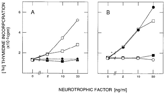
Quiescent gp140trk-expressing E25-48 cells (A) and gp145trkB-expressing Z52-17 cells (B) were incubated in DMEM containing 5 μg/ml insulin and the indicated amounts of NGF (open circles), baculovirus-derived BDNF(filled circles), baculovirus-derived NT-3 (open squares), or supernatant from wild-type baculovirus-infected Sf9 insect cells (filled squares). Incorporation of 3H-labeled thymidine into DNA was measured in duplicate samples 22 hr after stimulation as described in Experimental Procedures. Values are not corrected for DNA synthesis in untreated resting cultures.