
Fig. 1.
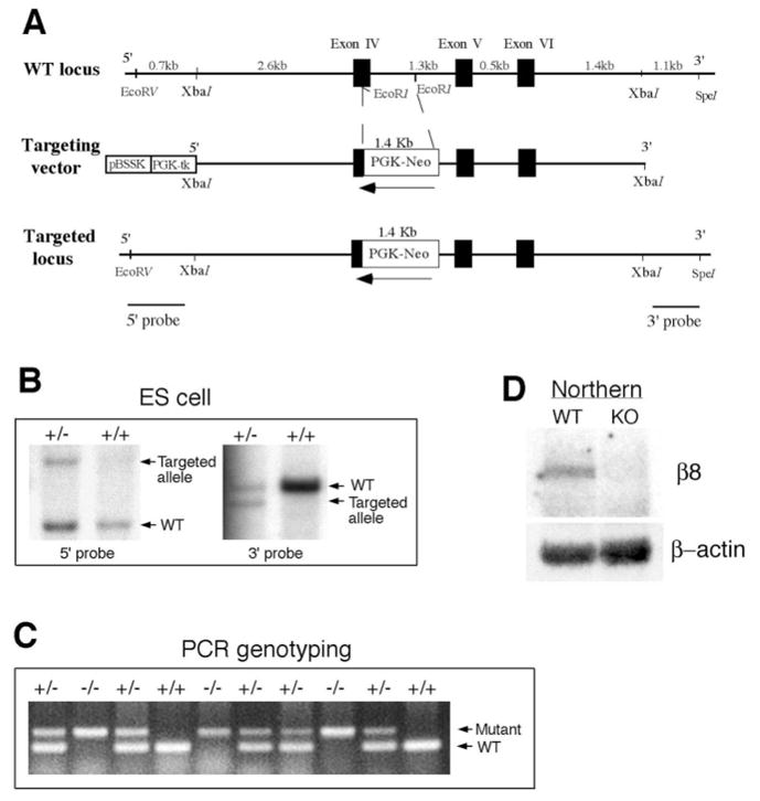
Generation of integrin β8-deficient mice. (A) Schematic drawing of integrin β8 genomic region encompassing Exons IV, V and VI in the wild type and mutant. Black boxes represent exons and white box represents the PGK-neor-cassette. Arrows indicate the transcriptional orientation of the cassette. 5′ and 3′ probes for Southern blot analyses are indicated. (B) Identification of ES clones containing a mutated β8 allele with 5′ and 3′ probes by Southern blot analyses. The wild-type and mutant alleles are labeled. (C) PCR analysis of genotypes from a heterozygous intercross. Mutant and wild-type amplification products are indicated. (D) Northern blot analysis showing the absence of integrin β8 transcripts in homozygous mutant mice. Comparable amounts of wild-type and mutant total RNA were loaded, as indicated by the presence of equal amounts of β-actin RNA. RT-PCR analysis further verified that no functional transcript is expressed in the mutant (data not shown).