Fig. 3.

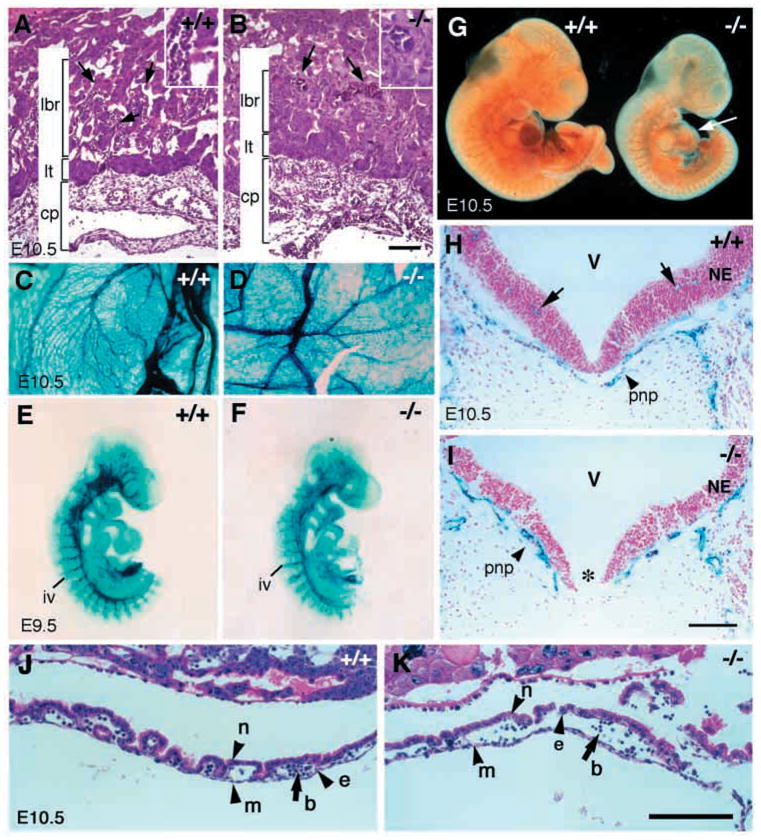
Angiogenesis defects in class A integrin β8-deficient mutants. (A,B) Hematoxylin and Eosin staining of transverse sections of placentas from an E10.5 wild type (A) and a mutant (B). While the chorionic plate (cp) and labyrinthine trophoblast layer (lt) are comparable in mutant and wild-type littermates, the labyrinthine layer (lbr) is reduced in the mutant. While the interdigitation of fetal blood vessels (A, arrows) and maternal blood vessels in wild-type embryos is elaborate, only a few fetal blood vessels have penetrated into the labyrinthine layer in the mutant (B, arrows). Inserts in A,B provide higher magnification photos of the vasculature. (C,D) Vasculature of E10.5 yolk sac in wild-type (C) and mutant (D) as depicted by X-gal staining of a Tie2:lacZ reporter gene in these mice. (E–G) Vascular patterns revealed by whole-mount staining of β-galactosidase activity in E9.5 embryos expressing the Tie2:lacZ reporter gene (E,F) and whole-mount immunohistochemistry with anti- PECAM antibody in E10.5 embryos (G). An E9.5 mutant embryo (E) shows no obvious abnormalities in vascular pattern compared with a wild-type littermate (F) (the tails of the embryos in both E and F were used for genotyping). E10.5 mutant embryos (G, right) have similar vascular patterns as wild-type littermates (G, left) except for reduced vasculature development in the heart (G, arrow). (H,I) X-gal staining of transverse sections of E10.5 neural tubes in wild-type (H) and mutant embryos (I) with the Tie2:lacZ reporter gene reveals the vasculature (blue) and cell nuclei labeled with Nuclear Fast Red (pink). While perineural plexuses are present in both the wild type and the mutant (H,I, arrowheads), there is no apparent penetration of vessels into the neural tube in the mutant when compared with the wild-type embryo (H, arrows). Note that the floor plate in the mutant (I, *) is absent. cp, chorionic plate; lt, labyrinthine trophoblast; lbr, labyrinthine layer; iv, intersomitic vessel; pnp, perineural plexus; V, ventricle; NE, neuroepithelium. (J,K) Hematoxylin and Eosin staining of E10.5 yolk sac showing presence of endothelial cells (e), mesothelial cells (m), blood cells (b) and endoderm cells (n) in both wild type (J) and mutant (K). Scale bars: 100 μm in A,B,H,I (500 μm in insets); 150 μm in J,K.
