
Fig. 5.
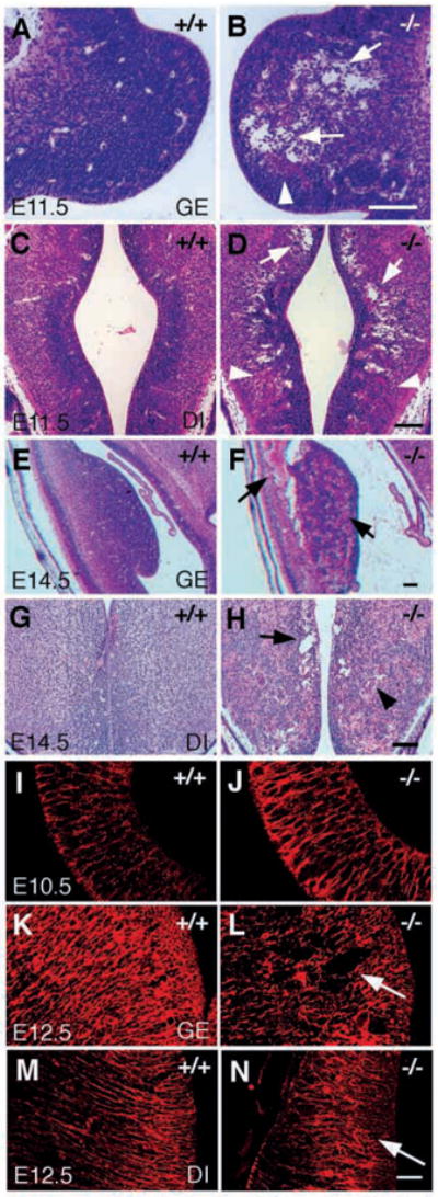
Abnormal cavitation and radial glial organization in the brains of class B integrin β8-deficient mutants. (A–H) Hematoxylin and Eosin staining of transverse sections of E11.5 brain (A–D) and E14.5 brain (E–H), showing abnormal cavitation in integrin β8-deficient mutant brains (B,D,F,H, arrows) compared with the wild-type littermates (A,C,E,G). Hemorrhage is visible in the ganglionic eminence (B, arrowhead) and diencephalon (D, arrowhead) of an E11.5 mutant brain and becomes much more severe in the E14.5 mutant brain (F, arrow; H, arrowhead). (I–N) Radial glial organization characterized by immunohistochemical staining using the RC2 antibody. Radial glial cells look grossly normal in an E10.5 mutant brain (J) compared with a wild-type littermate (I). However, they are apparently disorganized in the ganglionic eminence (L, arrow) and diencephalon (N, arrow) in the E12.5 mutant brain when compared with the same regions of E12.5 wild-type brains (K,M). GE, ganglionic eminence; DI, diencephalon. Scale bars: 100 μm in A–H; 50 μm in I–N.