
Fig. 6.
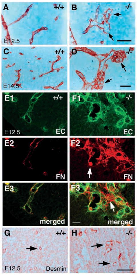
Abnormal brain capillary morphologies in class B integrin β8-deficient mutants. (A–D) Paraffin wax embedded sections of E12.5 (A,B) and E14.5 (C,D) brains stained with anti-laminin antibodies that show the abnormal morphologies of capillary vessels in the integrin β8-deficient mutant (B,D) when compared with a wild-type littermate (A,C). Discontinuous basement membranes are indicated by arrows (B). Aggregates of capillary vessels are visible (D, arrow). (E–F) Projected confocal images of E12.5 brain capillary vessels double labeled with anti-PECAM antibody (green) and anti- FN (red) antibodies. Capillary vessels in an integrin β8-deficient mutant (F1–3) exhibit irregular distended morphologies and are often conjoined when compared with those in wild-type littermates (E1–3). The basement membrane is discontinuous and a blood cell is captured at a potential hemorrhage site (F2, arrow) in the mutant. Note that a blood cell is present clearly outside of the capillaries, indicating hemorrhage in a nearby location (F3, arrow). (G,H) Pericytes recognized with anti-desmin are present and recruited to the capillaries in the brains of E12.5 wild-type (G, arrow) and mutant embryos (H, arrow). Scale bars: 50 μm in A–D; 20 μm in E,F; 100 μm in G,H.