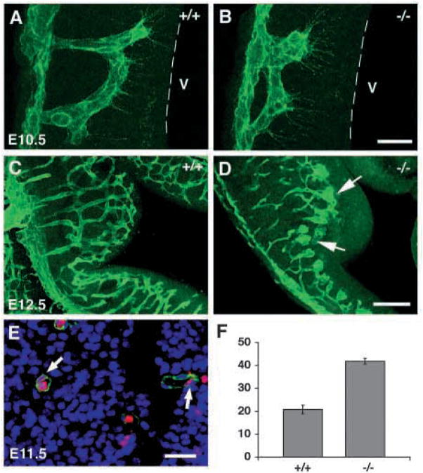
Fig. 8.
Abnormal capillary vascular patterning and endothelial cell hyperplasia in class B integrin β8-deficient brains. (A–D) Projected confocal images of brain capillary vessels stained with anti-PECAM in E10.5 (A,B) and E12.5 (C,D) embryos. Wild-type capillary vessels grow close to and couple near the ventricle (A, broken line marks boundary of ventricle, V). However, capillary vessels extend a shorter distance into the neuroepithelium and couple further away from the ventricle in the mutant (B). At E12.5, wild-type capillary vessels have branched and anastomosed to form an organized network (C); while in the mutant, capillary vessels show bulbous distentions and have not invaded regions of neuroepithelium immediately adjacent to the ventricle (D, arrows). (E) A brain capillary stained for isolectin BS (staining endothelial cell, green), DAPI (staining nuclei, blue) and BrdU (labeling proliferating cell, red, arrows). (F) The quantification of endothelial cell nuclei per vessel cross section in E11.5 wild-type and mutant brains. The percentage of BrdU-labeled endothelial cell nuclei in total endothelial cell nuclei scored is shown in the histogram (n=3). The error bars represent the s.e.m. Wild-type and mutant values are significantly different (P<0.005). Scale bars: 20 μm in A,B; 100 μm in C,D; 25 μm in E.

