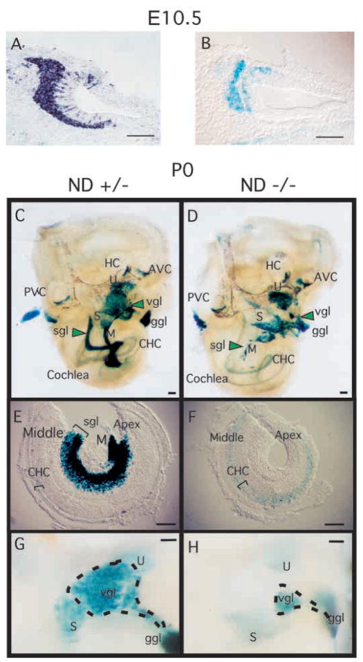
Fig. 2.
NeuroD is expressed in developing inner ear and is required for proper development of the inner ear sensory neurons. (A) In situ hybridization of E10.5 wild-type mouse embryo section using antisense NeuroD RNA as a probe and alkaline phosphatase reaction for visualization. (B) E10.5 NeuroD+/− mouse embryo section stained with X-gal. (C,D) Inner ear dissected from newborn (P0) NeuroD+/− (C) and NeuroD−/− mice (D), and stained with X-gal as whole mount. Anterior vertical canal (AVC), horizontal canal (HC), posterior vertical canal (PVC), saccule (S), utricle (U), vestibular ganglion (vgl), geniculate ganglion (ggl), spiral ganglion (sgl), modiolus (M) and cochlear hair cells (CHC) are indicated. In NeuroD−/− inner ear, the spiral ganglia (sgl) staining is absent and that of vestibular ganglia (vgl) is reduced and displaced compared with the control. (E,F) The apical and middle turn of the P0 cochleae are dissected and mounted after whole-mount staining with X-gal. The absence of spiral ganglia staining in NeuroD−/− inner ear is evident. Hair cells expressing lacZ are present in the NeuroD−/− cochleae. (G,H) Part of vestibular organ containing the vestibular ganglia, utricle and saccule is dissected out and mounted after whole-mount staining with X-gal to reveal the reduction in the size of the vestibular ganglia in NeuroD−/− mice. Scale bars: 100 μm.

