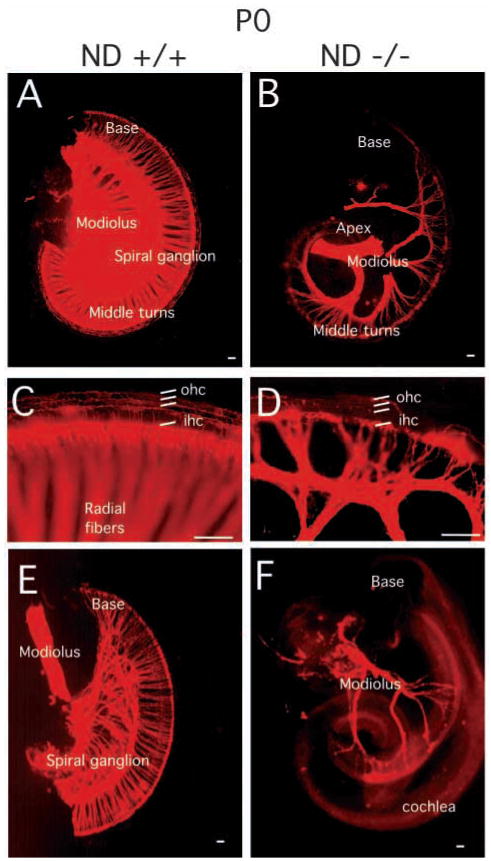
Fig. 3.
Innervation defects of the spiral ganglia in the NeuroD−/− mice revealed by DiI labeling. DiI labeling of the afferent nerve fibers (A–D) and efferent nerve fibers (E,F) innervating cochlear sensory epithelium at P0. The spiral ganglion tightly innervates the cochlear hair cells in sibling control animals (A). (B) In NeuroD−/− cochleae, only a few afferent fibers are present in the middle turn of the cochleae. (C,D) The higher magnification images showing the innervation to the inner and outer hair cells (ihc and ohc, respectively). (E) At P0, efferent fibers of sibling control animal reach the inner hair cells of the cochleae through out the entire epithelium. (F) In contrast, efferent fibers in the inner ear of NeuroD−/− mice are restricted to few fibers to the middle turn of the cochleae. NeuroD−/− efferent fibers fail to branch on their way from the modiolus to the cochlea. Scale bars: 100 μm.

