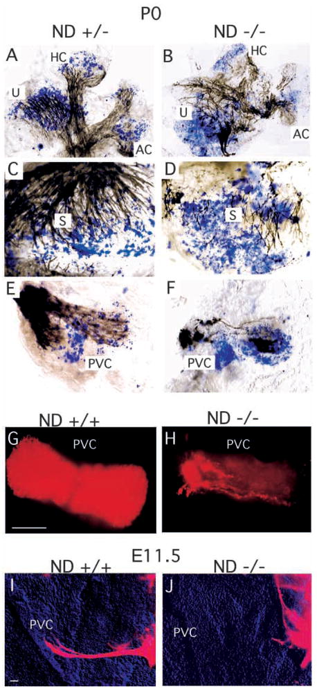
Fig. 4.
The pattern of innervation and of lacZ-expressing hair cells (HC: blue in A–F) in the P0 vestibular epithelia. (A–F) Dense innervation of all sensory epithelia in the NeuroD+/− animals (A,C,E) and the reduced density and partial absence of nerve fibers to parts of the sensory epithelium in NeuroD−/− mice (B,D,F) are shown in black. Also evident is disorganized fiber projection towards the sensory epithelia, as indicated by acetylated tubulin staining of the fibers. (G,H) DiI labeling of the afferent fibers to the PVC in control sibling (G) and NeuroD−/− (H) ears at P0 shows significant reduction in innervation into the PVC in NeuroD−/− mice. (I,J) DiI-labeling of the extending fibers at E11.5 indicates that fiber projection fails to initiate towards the PVC in NeuroD−/− mice. Scale bar in G, 100 μm for G,H; in I, 100 μm for I,J.

