Fig. 7.
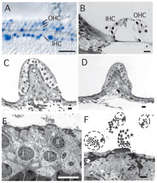
The inner ear hair cells develop and are maintained in the absence of innervation in NeuroD−/− mice. (A) Normally organized cochlear hair cells in NeuroD−/− mice are shown by X-gal staining. Many of the inner hair cells (IHC) and less of the outer hair cells (OHC) express various levels of lacZ. (B) Cochlear hair cells develop normally, even in 9-month-old animals in which the apical turn never received any innervation. (C–F) Electron micrographs of the posterior vertical canal (PVC) of a 9-month-old control (C) and NeuroD−/− mouse (D–F) show the absence of large calyxes, afferent innervation shown as empty space, around the Type I hair cells in NeuroD−/− mouse. The smaller size of the sensory epithelium and the absence of nerve fibers underneath the sensory epithelium are also evident. Despite the complete lack of innervation throughout embryonic and adult stages, hair cells are rather normal (D–F). Closer examination shows no nerve endings inside the sensory epithelium (D) but the presence of two types of hair cells (F): one type with stereocilia larger than kinocilia (asterisks), and the other with stereocilia the same diameter as kinocilia (# and circles). The Type I (I) and Type II (II) hair cells are marked in E. Note that the nuclei of the Type I cells are positioned deeper from the surface. In addition, the cuticular plate underneath the cilia coming out at the apex is thicker in the type I hair cells. The presence of two different types of hair cells suggests that development of the hair cells is independent of innervation. Scale bars: 100μm in A; 10 μm in B–E; 1 μm in F.

