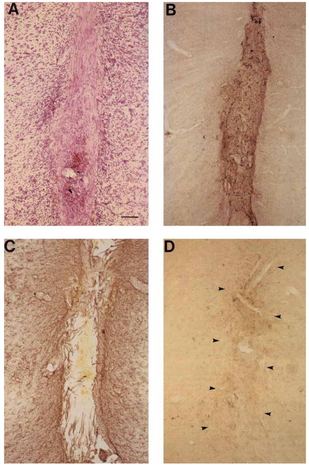
Fig. 8.
Coronal sections at the level of the substantia nigra show a typical FF/BDNF graft 6 weeks following implantation into the ventral mesencephalon. Sections taken through one FF/BDNF graft are stained with picrofuchsin, followed by a thionin counterstain (A), and immuno-stained with antibodies directed against FN (B), GFAP (C), and OX-42 (D). Arrowheads in D indicate graft borders. Scale bar = 100 μm.

