
Fig. 1.
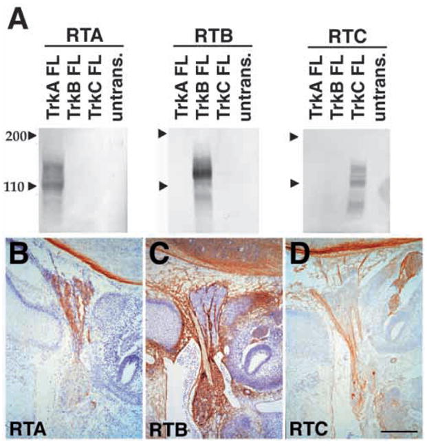
Specificity of Trk receptor antisera. (A) Western blot analysis of lysates of COS cells transiently transfected with full length rat TrkA, TrkB or TrkC, or of untransfected COS cells. Lysates were subjected to western blot analysis with RTA IgG (1 μg/ml; left panel), affinity purified RTB (1 μg/ml; center panel), or affinity purified RTC (1 μg/ml; right panel). Each antiserum recognizes protein bands of specific molecular weights in lysates of COS cells transfected with corresponding Trk receptor cDNA.
(B–D) Immunohistochemical staining of E13.5 IX-X ganglion complex. The intracranial superior jugular ganglion is at the top in these images and the extracranial nodose-petrosal complex is toward the bottom. RTA (B) identifies neurons in the superior jugular ganglion, but not the nodose-petrosal complex. RTB (C) recognizes neurons in the nodose-petrosal complex but not in the superior jugular ganglion and RTC (D) recognizes a small number of neurons in the nodose-petrosal complex. Scale bars (B–D) 200 μm.