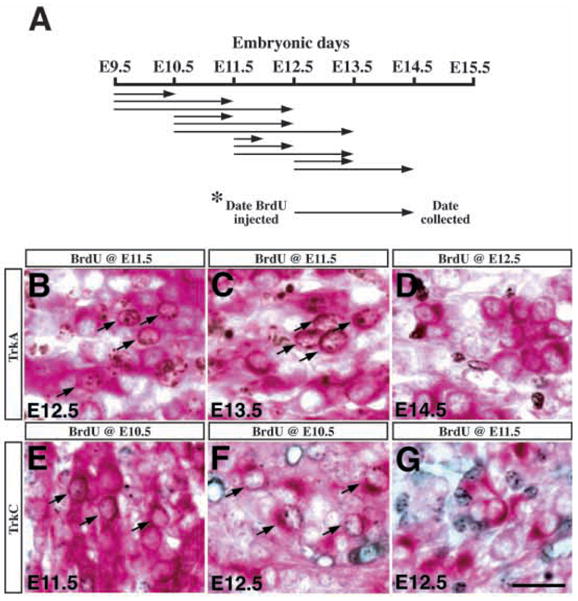
Fig. 6.

Neurons expressing different Trk receptors are generated at distinct time intervals during embryogenesis. (A) A schematic diagram indicating the experimental paradigm for birth-dating Trk-expressing neurons. The arrows designate the embryonic days when BrdU was injected intraperitoneally into the pregnant dams and the days when the embryos were collected for immunohistochemistry. (B–D) When pulse labeled with BrdU at E11.5, a significant number of TrkA-expressing neurons (red cytoplasmic staining) at E12.5 and E13.5 were immunoreactive for BrdU (brown nuclear staining) (arrows in B,C). However, when embryos received BrdU injections at E12.5 and were collected at either E13.5 or E14.5, there was no colabeling of BrdU and TrkA in the trigeminal neurons (D) (notice the complete separation of nuclear brown staining and red cytoplasmic staining). (E–G) Embryos labeled with BrdU at E10.5 showed colabeling of BrdU (black nuclear staining) and TrkC (red cytoplasmic staining) at E11.5 or E12.5 in the trigeminal neurons (arrows in E,F). However, in embryos injected with BrdU at E11.5 or E12.5, there was no colabeling of BrdU and TrkC in the trigeminal neurons (G). Scale bar, 20 μm.
