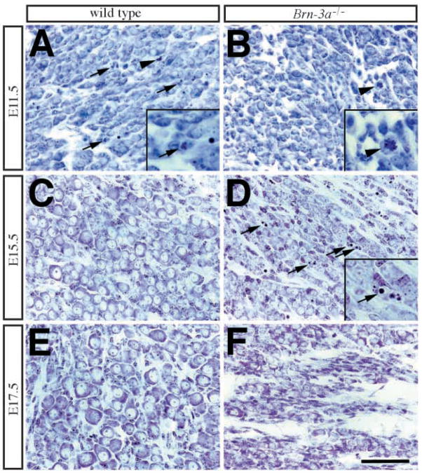
Fig. 3.
Morphological abnormalities in Brn-3a mutant ganglia at different embryonic stages. Cresyl violet (Nissl) stain of trigeminal ganglia from wild-type (WT) and Brn-3a mutant (Brn-3a−/−) mice at E11.5, E15.5, and E17.5. (A,B) At E11.5, cells in wildtype ganglia undergo apoptotic cell death (arrows) and mitosis (arrowhead), whereas in mutant ganglia, apoptotic profiles are fewer but mitosis is still present (arrowhead). (C,D) In contrast to the presence of well-developed sensory neurons with Nissl substance in wild-type ganglia (C), there are numerous apoptotic profiles in the mutant ganglia (D, arrows). The insets in A, B and D represent two-fold magnification of pyknotic nuclei (arrows in A,D) and mitosis (arrowhead in B). (E,F) By E17.5 and P0, the mutant ganglia contains much less neurons compared with that in the wild type. Scale bar, 50 μm.

