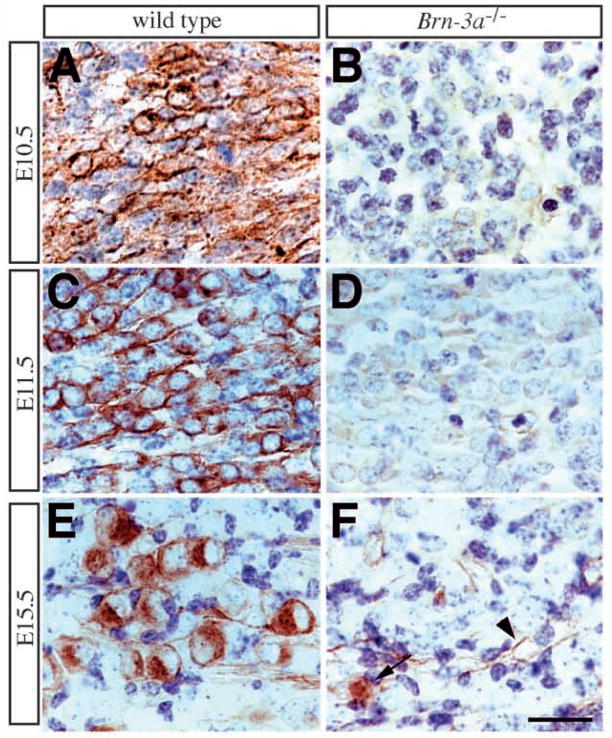
Fig. 5.
Failure to express TrkC in Brn-3a mutant ganglia. (A,C,E) Expression of TrkC receptor in wild-type ganglia shows a dynamic pattern during embryogenesis. TrkC expression is apparent in a small number of neurons at E10.5 (A). At E11.5, the number of TrkC-immunoreactive neurons becomes abundant and accounts for about 40–50% of neurons (C). However, this number decreases at E12.5 and remains unchanged after E13.5 (Table 1; Fig. 4D). At later stages, TrkC receptor is only present in a small number of neurons (E). (B,D,F) In contrast, there is essentially no TrkC receptor antigen detected at all stages. The light brown staining in mutant ganglia at E11.5 represents background color (D). Although rare TrkC-immunoreactive neurons can be identified at E15.5 (arrow, F), they constitute less than 10% of the wild-type TrkC neuron number (Table 1). TrkC expression in the vasculature is not affected in the mutant ganglia (arrowhead, F). Scale bar, 25 μm.

