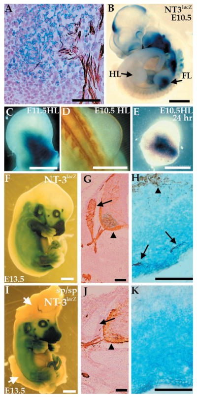
Fig. 1.
Expression of NT-3 in the limb does not depend on innervation. Expression of NT-3 was detected by a lacZ reporter gene stained with X-Gal (blue), and axons were visualized with a neurofilament antibody (NF, brown). (A) E11.5 NT-3lacZ embryo section shows colocalization of X-Gal staining with ends of sensory neuron projections. (B) E10.5 NT-3lacZ heterozygous embryo shows lacZ expression in forelimb (FL) but not hindlimb (HL). (C) E11.5 hindlimb stained for lacZ expression. (D) Dorsal view of an E10.5 embryo stained with NF. No axons are present in the hindlimb at this time. (E) E10.5 hindlimb cultured for 24 hours and stained for lacZ expression. (F to K) Expression of lacZ or NF (or both) in E13.5 NT-3lacZ embryos in a wild-type (F to H) or splotch background (I to K). Embryos were stained for lacZ expression (F and I). The splotch embryo exhibits severe defects in the cranial and lumbosacral region (arrows, I). No significant difference in lacZ expression in wild-type and mutant embryos is detected. (G and J) NF staining of caudal neural tube sections. DRGs (arrows) are absent in splotch mutants, although motor neurons (arrowheads) are present. (H and K) lacZ/NF costaining of the distal hindlimb. Sensory (arrows) but not motor (arrowhead) axons correlate with NT3 expression as detected by lacZ expression (H). (K) NT-3 expression is normal in the absence of NF staining in splotch mice. Bars, 50 μm (A, H, and K); 1 mm (B, F, and I); 0.5 mm (C to E); 100 μm (G and J).

