
Fig. 4.
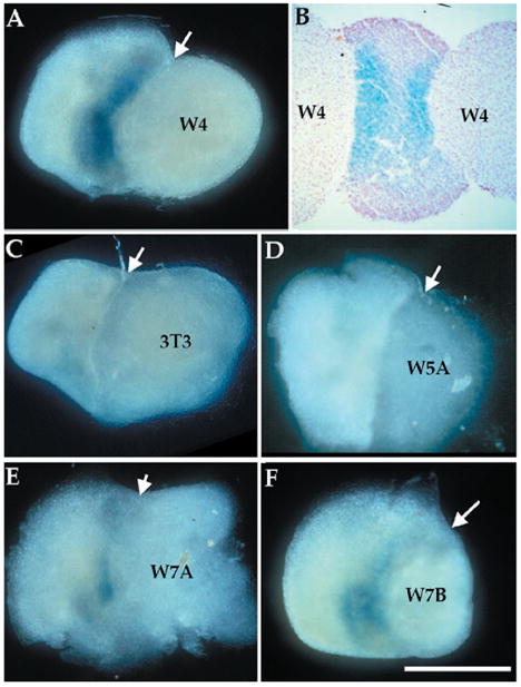
Wnt proteins are sufficient to induce NT-3 expression. Mesenchymal hindlimb tissue (without ectoderm) isolated from E10.5 NT-3lacZ heterozygous embryos was cocultured with aggregates of NIH 3T3–derived cell lines for 24 hours, after which they were stained with X-Gal. (A) Wnt-4–expressing NIH 3T3 cells induce NT-3 in limb mesenchyme. (B) A Wnt-4/limb/Wnt-4 explant stained and sectioned shows two blue stripes at the junction of the two tissues. (C) NIH 3T3 cells or (D) Wnt-5a– expressing cells do not induce NT-3, whereas (E) Wnt-7a– or (F) Wnt-7b–expressing cells induce NT-3 to a certain extent. Arrows point to junctions of limb mesenchyme and cell aggregates. Bar, 0.5 mm.