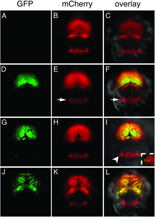
Figure 4.—
Distinct regions of the brain show differential silencing and reactivation. Corresponding images of GFP fluorescence (A, D, G, and J), mCherry fluorescence (B, E, H, and K), and an overlay of mCherry, GFP, and differential interference contrast (C, F, I, and L) from 3 dpf larvae. (A–C) c269 GFPoff larvae that are wild type or heterozygous for the dnmt1s872 mutation do not show GFP labeling. (D–F) c269 GFPoff larvae that are homozygous dnmt1s872 mutants show GFP labeling in the midbrain and forebrain. Despite transactivation of mCherry, GFP labeling is not observed in r5–6 (arrow in E and F). (G–I) The homozygous dnmt1s872 mutant with gfp reactivation in a small number of bilateral cells in r5–6. The insert in I is an enlargement of reactivated cells indicated by the arrowhead. (J–L) Larvae from a c269 GFPhigh adult mated to wild type. Labeling of GFP is present in the forebrain and midbrain, but not in r5–6 mCherry-positive cells.

