Figure 1.—
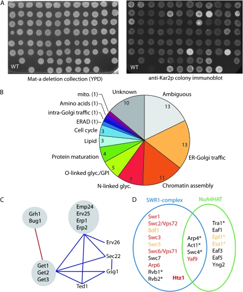
Overview of the Kar2p retention screen and categorization of mutants. (A) The haploid deletion collection was grown on YPD (left) and secreted Kar2p was quantified using fluorescent secondary antibodies (right). Colonies secreting twofold more Kar2p than WT cells were selected for further characterization. (B) Functional distribution of Kar2p secretors and the number of genes isolated in each category. (C) Genetic interactions among Kar2p secretors with annotated functions in ER-Golgi traffic. Components that have been isolated as physical complexes are shown in gray. Aggravating interactions are shown in blue, alleviating interactions in red. (D) Subunit architecture of the SWR1 and NuA4 complexes. Kar2p secretors are shown in red, components that showed a less significant Kar2p secretion phenotype are shown in orange, essential genes are annotated with an asterisk. Adapted from Kobor et al. (2004).

