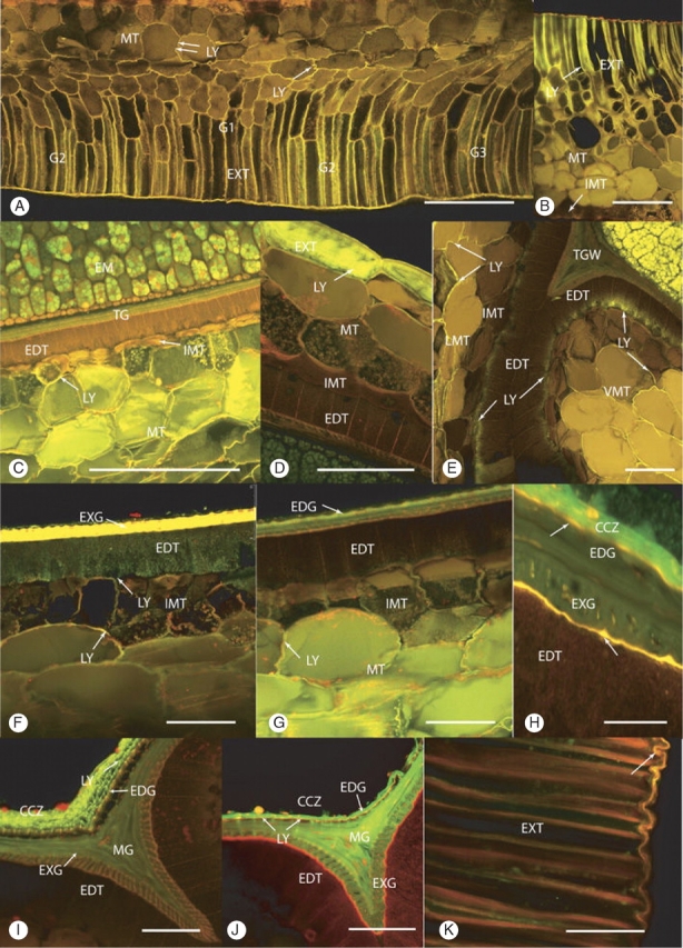
Fig. 3.

(A–C) Cross-sections showing the movement of Lucifer Yellow (LY) into the seed following 24-h immersion. (A) Heat- and smoke-treated seed, zone 2, ventral. LY had penetrated up to the innermost layer of the mesotesta (double arrows) in the G1 and G2 regions. There are two to three layers of columnar-shaped cells in the G1 region. These cells are probably exotestal in origin. The intensity of LY-induced fluorescence is greatest in the columnar exotestal cells, decreasing in the outer squat-shaped mesotestal cells (single arrow) and is weakest further inwards (double arrows). Scale bar = 250 µm. (B) Control seed, ventral. LY-induced fluorescence was found halfway into the mesotestal layer of the testa. Scale bar = 180 µm. (C) Heat- and smoke-treated seed, zone 3, ventral, merged image. LY had penetrated through the mesotesta reaching but not entering the innermost mesotestal layer (IMT). Embryo (EM), endotesta (EDT), tegmen (TG), mesotesta (MT). Scale bar = 250 µm. (D–G) Cross-sections through whole, intact seeds that were immersed in Lucifer Yellow for 48 h. (D) Control seed, zone 3, dorsal. LY has diffused beyond the exotesta into the outer mesotestal layer. Endotesta (EDT), exotesta (EXT), innermost mesotestal layer (IMT), mesotesta (MT). Scale bar = 50 µm. (E) Control seed, zone 2, ventral G3. LY penetrated through the lateral mesotesta (LMT) into the outer part of the endotesta cells, forming the neck region of the endotesta wing (WEDT). LY has also diffused through the curved area of the ventral groove into the endotesta. Endotesta (EDT), innermost mesotestal layer (IMT), tegmal wedge (TGW). Scale bar = 100 µm. (F) Smoke-treated seed, zone 3, ventral, G1. LY penetrated through the mesotesta and into the endotesta (EDT). Innermost mesotestal layer (IMT), exotegmen (EXG). Scale bar = 50 µm. (G) Smoke-treated seed, zone 3, ventral, G1. LY has diffused through the mesotesta (MT) and entered the radial walls of the innermost layer of the mesotesta (IMT), but has not progressed into the abutting endotesta (EDT). Scale bar = 50 µm. (H–K) Cross-section of seeds that were halved, the embryo carefully removed and Lucifer Yellow applied to the cavity for 24 h of outward diffusion. (H) Heat- and smoke-treated seed, zone 3, G1, counterstained with Nile Red. LY was confined to the crushed cell zone (CCZ). The two inner cuticles (arrows) fluoresced gold. Endotegmen (EDG), exotegmen (EXG), endotesta (EDT). Scale bar = 25 µm. (I) Control seed, zone 3, tegmal wedge. LY is retained in the crushed cell zone (CCZ) with no passage through the inner cuticles. Endotegmen (EDG), exotegmen (EXG), endotesta (EDT), mesotegmen (MG). Scale bar = 50 µm. (J) Heat- and smoke-treated seed counterstained with Nile Red, zone 3, tegmal wedge. LY was present in the crushed cell zone (CCZ) and within the tegmal layers. Stronger lines of LY indicated where the dye has moved into the tegmen via cuticular pores (arrows). A break in the CCZ and the inner cuticle also permitted movement of the dye into the tegmen. The thicker inner cuticle (large arrow) between the endotesta and the exotegmen fluoresces red rather than orange. Endotesta (EDT), endotegmen (EDG), mesotegmen (MTG), exotegmen Scale bar = 50 µm. (K) Control seed, unfixed, zone 1, ventral, G1, stained with Nile Red. The suberized walls of the exotestal (EXT) cells strongly fluoresce red. The overlying outer cuticle (arrow) fluoresce orange gold. Scale bar = 50 µm.
