
Fig. 1.
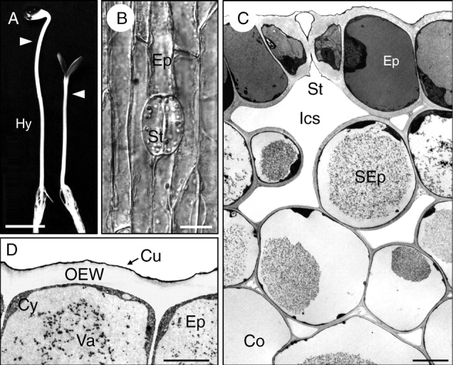
Axial plant organs and the growth-limiting outer cell layers. (A) Photograph of sunflower (Helianthus annuus) seedlings that were either grown for 4 d in darkness (left) or for 3 d in darkness and 1 d in continuous white light (irradiated plant, right). (B) Light micrograph of the epidermis in the sub-apical region of the hypocotyl (arrowheads in A) of an irradiated seedling. (C, D) Electron micrographs of transverse sections through the peripheral four cell layers in the sub-apical region of a 4-d-old irradiated seedling (C) and the epidermis (D). Co, cortex; Cu, cuticle; Cy, cytoplasm; Ep, epidermis; Hy, hypocotyl; Ics, intracellular space; OEW, outer epidermal wall; SEp, sub-epidermis; St, stoma; Va, vacuole. Scale bars: (A) 1 cm, (B) 20 µm, (C) 10 µm, (D) 2 µm.