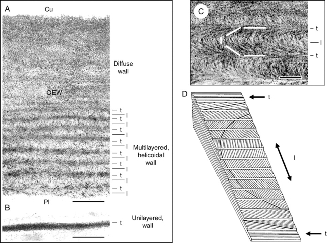
Fig. 2.

Cellulose architecture of outer and internal walls of a 4-d-old sunflower seedling that was grown in darkness and white light (see Fig. 1A). Transmission electron micrographs of oblique transverse sections through the outer epidermal wall (A, C) and a wall in the pith (B). The cellulose microfibrils were stained with periodic acid–thiocarbohydrazide silver protein (PATAg) as described by Hodick and Kutschera (1992). A model of a helicoid (D) shows parallel cellulose microfibril layers that are packed with a constant rotation (adapted from Mulder and Emons, 2001). l, longitudinally orientated; t, transversely orientated cellulose layers with respect to the axis of cell elongation; Cu, position of the cuticle; OEW, outer epidermal wall; Pl, position of the plasma membrane. Scale bars = 0·5 µm (A, B), 0·1 µm (C).
