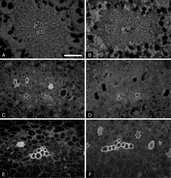
Fig. 1.

Microphotographs of cross-sections of soybean seedlings grown in 1 g on Earth (A, C, E) and in microgravity (B, D, F): cotyledons (A, B), hypocotyl hook (C, D) and hypocotyl (E, F). Vessel walls appear fluorescent because of the presence of lignin. Single principal veins are surrounded by parenchyma cells in cotyledons (A, B). Single poles of xylem in the stele of hypocotyl zones are shown (C, D, E, F). Scale bar = 50 µm.
