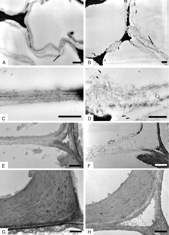
Fig. 4.

Transmission electron microphotographs of thin sections of developing vessel walls of soybean seedlings grown in 1 g on Earth (A, C, E, G) and in microgravity (B, D, F, H). Periodic acid–thiocarbohydrazide–silver-proteinate staining. Cellulose microfibrils lie parallel to each other and are arranged in lamellae in cotyledons (A, C) and hypocotyl (E) of seedlings grown in 1 g. Microfibrils are scattered and are not easily arranged in thicker bundles in cotyledons (B, D) and hypocotyl (F) of seedlings developed in microgravity. Vessel walls with complete deposition of secondary wall show ordered arrangement in both 1 g (G) and microgravity (H). Long arrows = lamellae; short arrows = microfibrils. Scale bars = 0·5 µm.
