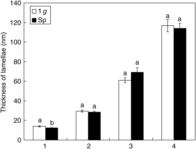
Fig. 5.
Arrangement of cellulose microfibrils into thicker bundles in vessel walls of soybean seedlings. Size of lamellae resulting from the progressive arrangement of microfibrils in seedlings developed in normal gravity conditions (1 g) and in microgravity (Sp). The numbers from 1 to 4 label the lamellae of increasing thickness starting from the thinnest detectable. Mean values and standard errors are shown. Different letters correspond to significantly different values after using LSD and Student–Newman–Keuls coefficients for multiple comparison tests (P < 0·05).

