
Fig. 2.
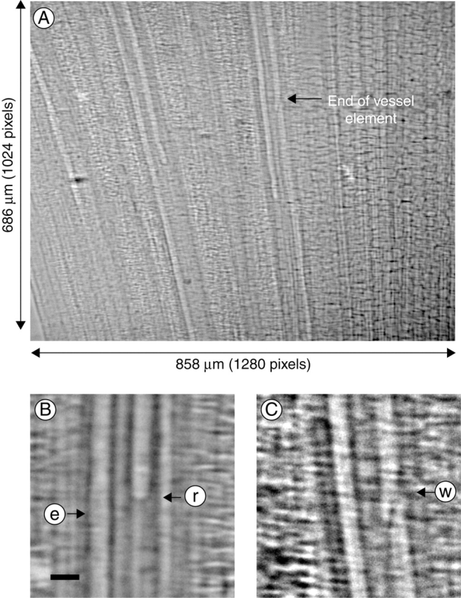
A typical X-ray image of the xylem vessels within a bamboo leaf cut in air, allowed to dry briefly and rehydrated. Image A is an X-ray image of the leaf, while images B and C are micrographic sections showing the meniscus (between water and air) in a xylem vessel and the end wall of the xylem vessel element. In B, arrows e and r show an empty and refilling xylem vessel, respectively. In C, the end wall between two adjacent vessel elements is shown by arrow w. Scale bars = 20 µm.