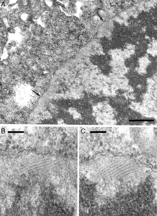
Fig. 8.

Electron micrographs of pollen mother cells of Triticum aesticum ‘Chinese Spring’ at premeiotic interphase showing (A) two bundles of fibrillar material (FM; arrowed) apparently linking chromatin to nuclear membrane; (B, C) FM from (A) at higher magnification showing bundles composed of microfibres (image taken from Bennett et al., 1979). Scale bars: (A) = 0·5 µm; (B, C) = 100 nm.
