
Fig. 1.
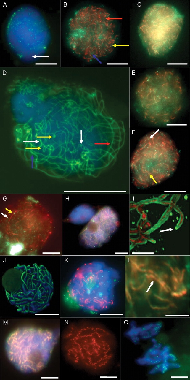
(A) Early leptotene in spring rye, showing Asy1 foci (green), and an area in the nucleus with relatively few foci (white arrow). The nucleus is counter-stained with DAPI (blue). (B) Mid-leptotene in spring rye showing Asy1 (red), Spo11 (green), a pair of Spo11 foci (red arrow), a Spo11 focus co-localized with an Asy1 core (yellow arrow), and a chain of three Spo11 foci (blue arrow). (C) Mid-leptotene in spring rye showing Asy1 (red) and Rad51 (green). (D) Zygotene in spring rye showing Asy1 (green), fold-back loops or aligned telomeres (yellow arrows), distant (red arrow) and close (blue arrow) alignment of Asy1 cores, and synapsed/paired regions (white arrows). The nucleus is counterstained with DAPI (blue). (E) Mid-zygotene in spring rye showing Asy1 (red) and Rad51 (green). (F) Mid-zygotene in spring rye showing Asy1 (red), Spo11 (green), a Spo11 focus co-localized with an Asy1 core (white arrow) and a Spo11 focus lying between two aligned Asy1 cores (yellow arrow). (G) Pachytene in spring rye showing Asy1 (red), Spo11 (green), a Spo11 focus adjacent to (yellow arrow) and co-localized with (white arrow) an Asy1 core. (H) Pachytene in spring rye showing complete co-localization of Asy1 (green) and Zyp1C (red). (I) Detail from a projection of an optical stack through pachytene in spring rye showing Asy1 (green), Zyp1C (red) and a cap of Asy1 (arrow). (J) Projection of an optical stack through pachytene of mutant sy1 showing Asy1 (green) and DAPI counterstain (blue). (K) Diplotene in spring rye showing Asy1 (red) and Spo11 (green). (L) Detail of diplotene in spring rye showing co-alignment (arrow) of desynapsed cores of Asy1 (red) and Zyp1 N (green). (M) Diplotene in spring rye showing widespread co-alignment (yellow) of Asy1 (red) and Zyp1 N (green). (N) Mid-diplotene in Sy10 showing spiralling of Asy1 cores only (red). (O) Late diakinesis showing fragments of Asy1 (green). All bars represent 10 µm, except those of (I) and (L) which represent 5 µm.