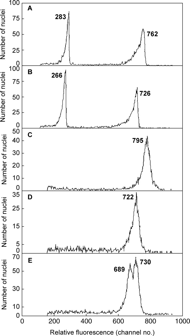
Fig. 2.

Relative DNA staining in 2C nuclei of Pisum sativum co-chopped with green leaf (A) or red bract tissue (B) of Euphorbia pulcherrima and of Pisum sativum suspended in Galbraith buffer with no added anthocyanin (C), or with 200 µm of added cyanidin-3-rutinoside (D), or (E) after stained nuclei of C and D were mixed. The mean relative absorbance for nuclei in each peak is given.
