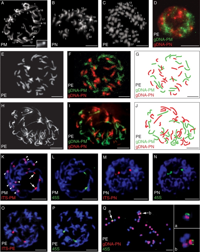
Fig. 2.

(A–C) DAPI-stained root tip prophases/metaphases of (A) Primula mistassinica showing 2n = 18 chromosomes, (B) P. nutans showing 2n = 22 chromosomes, and (C) P. egaliksensis showing 2n = 40 chromosomes; the arrowhead in (A) points to the heterochromatic knob shown in the inset. (D) GISH to interphase nuclei of P. egaliksensis using gDNA from P. mistassinica (green) and P. nutans (red). (E–J) Root tip prophases/metaphases of P. egaliksensis stained with DAPI (E, H), and resulting GISH signal (F, G, I, J) following in situ hybridization of gDNA from P. mistassinica (green) and P. nutans (red). (K–P) FISH localization of ITS (red) and 45S rDNA (green) loci on root tip prophases of (K, L) P. mistassinica, (M, N) P. nutans, and (O, P) P. egaliksensis counterstained with DAPI; in (K), arrows point to cross-hybridization of ITS and 45S rDNA probes, and arrowheads highlight heterochromatic knobs. (Q) Root-tip metaphase of P. egaliksensis counterstained with DAPI after GISH with gDNA of P. nutans (red) and FISH with 45S rDNA probes (green); arrows (a, b) point to the two 45S rDNA-bearing chromosomes shown in the insets. Scale bar = 10 µm. PE, P. egaliksensis; PM, P. mistassinica; PN, P. nutans.
