Fig. 1.

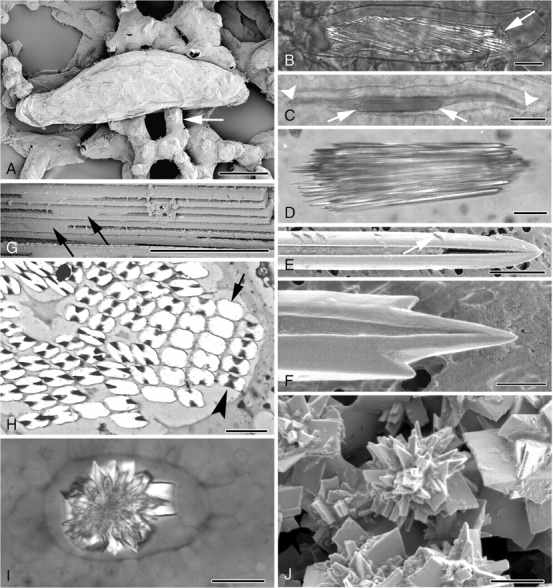
(A–J) Light and electron micrographs of cell inclusions in Amorphophallus. (A) Elongated raphide idioblast in leaf tissue of A. titanum. Arrow points to adjacent mesophyll cell. (B) Polarized-light image of a raphide idioblast of A. variabilis. Crystals are contained within the cell vacuole (arrowed). (C) Raphide bundle (arrowed) in A. konjac, significantly shorter than the length of the cell vacuole, marked by arrowheads. (D) Individual raphide crystals in A. konjac taper to pointed tips. (E) Barbs (arrowed) can be seen along the length of a raphide of A. salmoneus. One of the two characteristic grooves running the length of the crystal can be seen. (F) Additional points are present on either side of the pointed end of a raphide of A. salmoneus. (G) The crystal grooves disappear around the mid-point of each needle, as here in A. konjac. (H) Transverse section through a raphide bundle of A. bulbifer. At mid-point of crystals absence of groove results in rectangular crystal outlines (arrow), whereas at other points along the crystals the outlines are characteristically H-shaped (arrowhead). (I) Polarized-light image of a druse of A. salmoneus. (J) Druse in A. salmoneus, numerous crystal faces amalgamated together around a central region. Scale bars: (A, C) = 50 µm; (B, D, I) = 20 µm; (E, H) = 2 µm; (F, J) = 1 µm; (G) = 5 µm.
