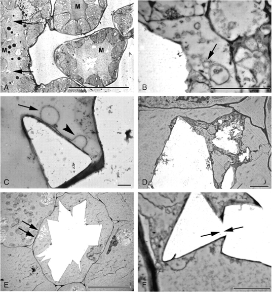
Fig. 8.

Druse development, mid- and late stages in Amorphophallus titanum (A) and A. krausei (B–F). (A) Multiple druses (arrowed) developing in an elongated idioblast. Note stained mucilage (M) in vacuoles. (B) Numerous vesicles (arrowed) in close proximity to developing druse. Jagged edge of druse suggesting crystal deposition in steps or layers. (C) Membranous sheath around crystal face, often apparently looped (arrowhead). Vesicle (arrowed) in contact with sheath. (D) Crystal deposition occurring in several areas within the vacuole. (E) Mature druse, filling cell. Crystal deposition also seen (arrowed) in another part of the vacuole. (F) Crystal faces with differing orientations are interlocked (arrowed). Scale bars: (A, E) = 20 µm, (B) = 1 µm; (C) = 400 nm; (D) = 4 µm; (F) = 2 µm.
