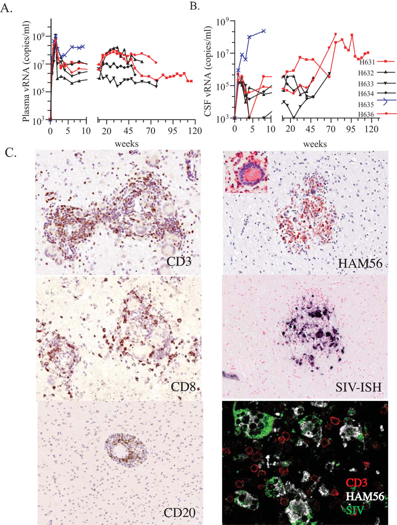
Fig. 1.
Clinical and pathological features. (A) Plasma and (B) CSF viral RNA levels. Samples were obtained at sequential time points post inoculation. Y axis represents viral RNA load (copies/ml) and the X axis represents weeks post inoculation. Macaques H631 (square symbol) and H636 (circle symbol) are shown in red and the rapid progressor macaque H635 is shown in blue. (C) SIV-specific in situ hybridization and immunohistochemistry of infiltrating cell populations in the brain of conventional progressor macaques with SIVE, H631 and H636. Prominent perivascular mononuclear infiltrates stained specifically with antibodies to CD3 (H631), CD8 (H631), and CD20 (H636) on the left identified by DAB substrate (brown). The right panels show the infiltration of macrophages as evident by staining with HAM56 in red (H636), with an inset of a characteristic HAM56+ multinucleated giant cell. A serial section shows the expression of SIV RNA in these infiltrating cells in blue. The bottom right panel shows confocal microscopy of the brain of H636 showing that SIV-expressing cells (green), co-expressed HAM56 (white) consistent with their identification as macrophages although uninfected CD3+ T cells (red) were also present.

