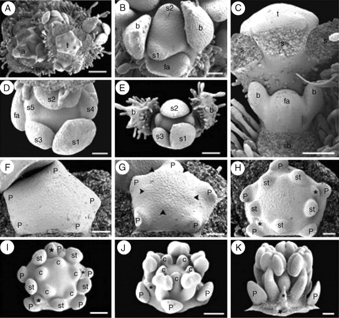
Fig. 9.

Suriana maritima floral ontogeny. SEM micrographs. (A) Young flowering branch with lateral floral bud initiation. (B) Polar view of floral bud during sepal initiation flanked by a younger floral apex (bottom). (C) Lateral view of a partial inflorescence with lateral floral apex immediately after bracteole initiation; sepals of terminal flower were removed. (D) Polar view of floral bud after helical sepal initiation. (E) Polar view of a young dichasial partial florescence; terminal flower surrounded by bracts subtending young floral buds. (F) Floral bud during petal initiation. (G) Floral bud during simultaneous initiation of antesepalous stamen primordia (arrowheads). (H) Floral bud with antesepalous stamen primordia alternating with two staminode primordia (asterisks). (I) Floral bud after carpel initiation; carpels are initiated opposite staminodes (asterisks) and petals. (J) Oblique view of floral bud with basifixed anthers, staminode (asterisk) and concave carpels at the top of enlarged gynophore. (K) Lateral view of floral bud with stamens, staminode (asterisk) and floral base becoming hairy; petal base is pointed (arrow). a, branch apex; b, bracteoles; c, carpel; fa, floral apex; p, petal; s, sepal; s1–s5, order of sepal initiation; sb, subtending bract position; st, stamen; t, terminal flower. Scale bars: A = 200 µm; B, D, F–H = 50 µm; C, E, I = 100 µm; J, K = 150 µm.
