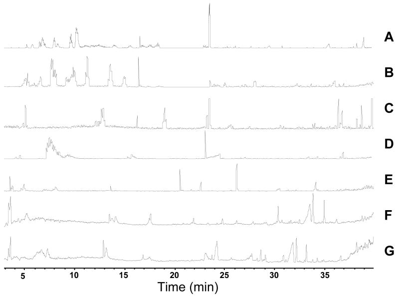
Figure 5.
Base peak chromatograms from 1D and 2D separation of small molecules from E. coli. RPLC was performed using 30 cm × 25 μm I.D. column. 2D was performed by off-line fraction collection in 10 minute step intervals from SAX capillary column. [Brackets] indicate the average number of small molecules detected. A:1-D RPLC-MS [170] B-G 2-D SAX-RPLC B: 5 mM NH4HCO2 fraction [79] C. 100 mM NH4HCO2 fraction [46] D. 200 mM NH4HCO2 fraction [45] E. 300 mM NH4HCO2 fraction [38] F. 400 mM NH4HCO2 fraction [19] G. 500 mM NH4HCO2 fraction [16]

