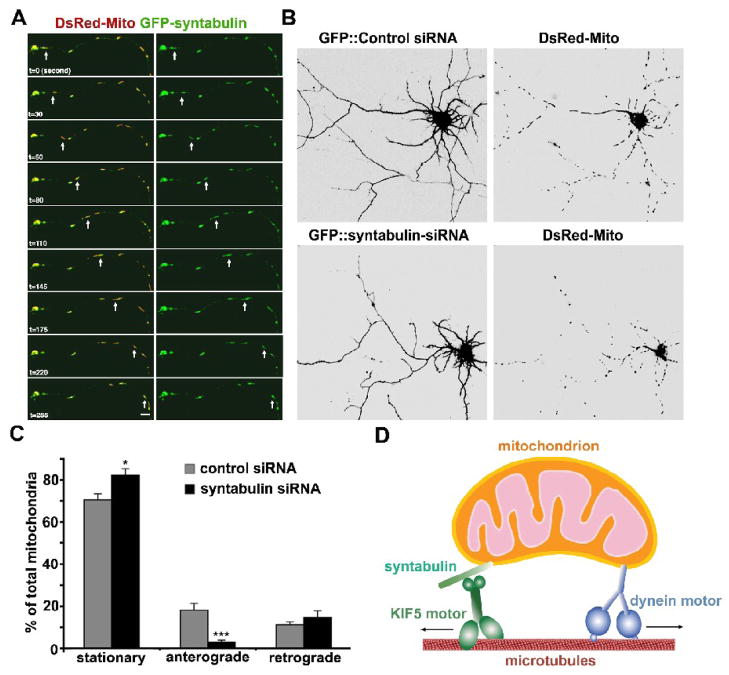
Fig. 2.
Syntabulin serves as a KIF5-adaptor and mediates anterograde transport of mitochondria along neuronal processes. (A) Syntabulin and mitochondria co-localize and co-migrate along axonal process. Cultured hippocampal neurons (DIV 6) were co-transfected with GFP-syntabulin (green) and DsRed-mito (DsRed2-tagged mitochondrial targeting sequence of cytochrome c oxidase, red) and the selected axonal process was time-lapse imaged 18 hours after transfection. Syntabulin-associated mitochondria move along the axonal processes. The arrows point at a moving mitochondrion associated with EGFP-syntabulin, which migrated anterogradely along the axon toward the growth cone at an average velocity of approximately 0.3–0.4 μm/sec. Images were collected every 5 seconds. Scale bar in panel B equals 10 μm. (B) Knockdown of syntabulin expresson reduces mitochondrial density in the neuronal processes of cultured hippocampal neurons. Hippocampal neurons (DIV4 or 5) were co-transfected with DsRed-mito and syntabulin-targeted siRNA or control siRNA. Mitochondrial distribution was examined by imaging GFP fluorescence (left panels) and DsRed-mito (right panels) in the neurons 4 or 5 days after transfection. Note that in the processes of neurons expressing syntabulin-siRNA, an abnormally lower density of mitochondria was observed relative to that of the neurons transfected with control siRNA. Scale bars, 10 μm. (C) Syntabulin loss-of-function impairs anterograde movement of mitochondria along axonal processes. The motility of DsRed-mito-labeled mitochondria was examined in live hippocampal neurons at DIV9-10 after tranfection with siRNAs. The direction of net movement for each mitochondrial organelle along axonal processes was determined during the same time window (15 min) of time-lapse imaging and the relative percentages of stationary, net anterograde, or net retrograde events were calculated. Knockdown of syntabulin inhibits anterograde but not retrograde movement of mitochondria along axonal processes. Of a total of 259 mitochondrial clusters from 11 cells transfected with the control-siRNA, one-third of the mitochondrial organelles in the axons are mobile (29.6 ± 3.0%), which moved either anterogradely (18.4 ± 2.9%) or retrogradely (11.2 ± 1.6%). Knockdown of syntabulin (total 383 mitochondria from 17 cells) exhibited a marked reduction in anterograde transport (***p<0.001), a slight but significant increase in stationary mitochondria (*p<0.02), and no significant change in retrograde movement of mitochondria (p=0.4). (D) Schematic diagram for the proposed role of syntabulin. Syntabulin serves as an adaptor in linking the KIF5 motor to mitochondria and in mediating anterograde transport of mitochondria in axons. (The images and quantification data are adapted with permission from Qian Cai, Claudia Gerwin, and Zu-Hang Sheng. Syntabulin-mediated anterograde transport of mitochondria along the neuronal processes. Journal of Cell Biology 170, 959–969. 2005).

