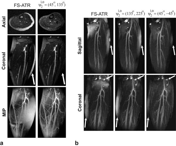
Figure 4.
Comparison of FS-ATR and the proposed method at two different field strengths: a: 1.5 T, b: 3 T. a: Coronal and axial slices for the FS-ATR and acquisitions at 1.5 T are displayed along with the corresponding MIPs. The arrows point to the region where the proposed method achieves better fat suppression than the FS-ATR method. Improved fat suppression of the proposed method results in superior depiction of the vasculature in the MIPs. However, regions with visible residual fat signal still exist in the images produced with the proposed method as a result of the remnant stop-band signal. b: Sagittal and coronal thin slab MIPs of the calf acquisitions at 3 T are displayed for the FS-ATR (ψ2 = 180°), and methods. The combination achieves better fat suppression than FS-ATR. However, the range of off-resonant frequency variation limits the amount of fat suppression. The combination achieves the highest level of fat suppression due to its broad stop-band.

