
Fig. 3.
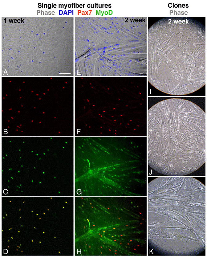
Immunofluorescent and phase images depicting myogenesis in cultures emanating from single EDL myofibers and in live clones of satellite cells that were dissociated from EDL myofibers. Myofibers from a young mouse were cultured for 1- or 2-weeks (A–D and E–H, respectively) and then double-labeled for Pax7 and MyoD. (A, E) Merged phase and DAPI images; (B, F) Pax7; (C, G) MyoD; (D, H) merged Pax7 and MyoD images demonstrating that all cells in the earlier time point co-express Pax7 and MyoD but at the later time point only residual cells are positive for both antigens. Clones were derived from EDL myofibers of young (I), old (J) and senile (K) mice. Bar, 100 μm.