
Fig. 5.
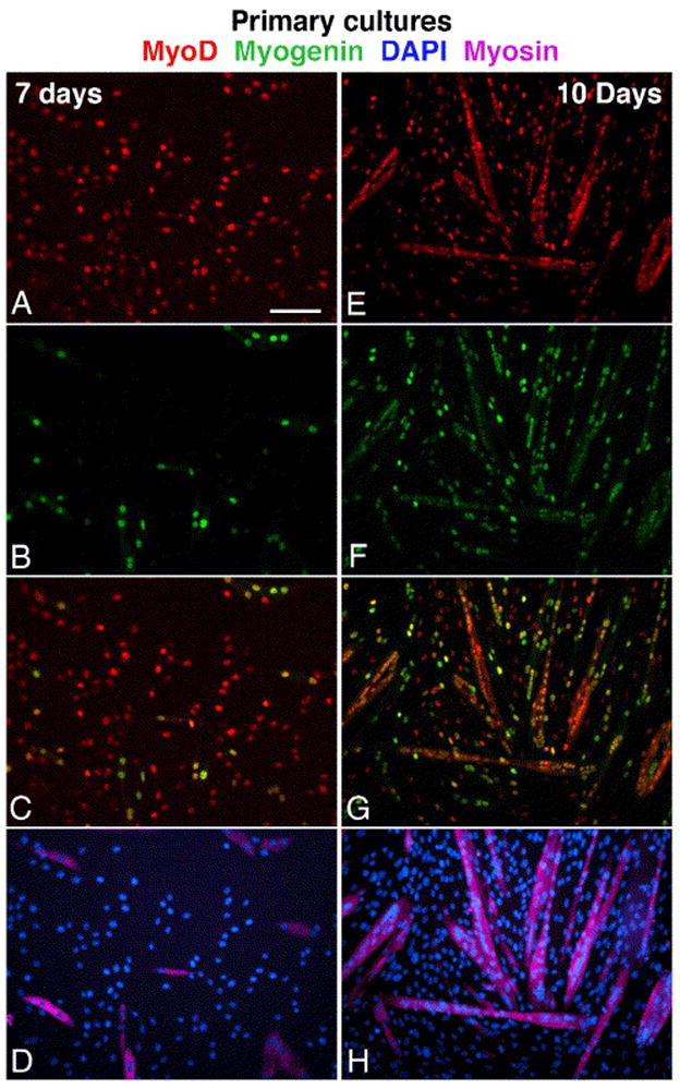
Parallel immunofluorescent images depicting myogenesis in primary cultures prepared from the tibialis anterior and gastrocnemius muscles of senile mouse. Cultures shown were fixed on day 7 (A–D) and day 10 (E–H). Cultures were triple-immunolabled for MyoD (A, E), myogenin (B, F) and sarcomeric myosin (D, H; these images merged with parallel DAPI stain). Merged images of MyoD and myogenin are also shown (C–G). The same immunostaining patterns were observed in cultures from young mice, but with an earlier onset of differentiation. Bar, 100 μm.