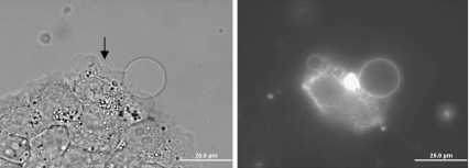
Fig. 6.
Irradiated cells externalize PS lipids on all the exposed cell membranes. Bright field and fluorescent micrographs of KB cells surrounded by Annexin V-Alexa Fluor 488 and binding buffer at 37°C are shown. The single irradiated cell (as indicated by the arrow) is atop a group of about eight other cells also within view. The control cells show no PS externalization. The irradiation focal spot shows brightest on the fluorescence image

