Fig 1.
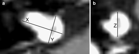
a, b Contrast-enhanced T1-weighted image with a vestibular schwannoma in the cerebellopontine angle (CPA) on the right side. a Measurements in the axial plane: X is the maximum mediolateral, and Y the maximum anteroposterior dimension; b in the coronal plane, the Z demonstrates the maximum craniocaudal dimension

