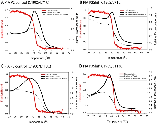
Figure 5. Excimer fluorescence of PIA-labeled and light scattering of unlabeled P2 and P3 mutants.
A. P2 control (C190S/L71C) B. P2Shift C190S/L71C C. P3 control (C190S/L113C). D. P3Shift C190S/L113C. Light scattering (red solid line and left axis with red label) was conducted with unlabeled tropomyosin due to interference from excimer peak in light scattering experiments with labeled tropomyosin. Excimer fluorescence (right axis) in the presence of actin (black solid line) and absence of actin (black dotted lines). Buffer conditions and the experimental procedure are as described for Figure 4. The P2Shift and P3Shift mutations locally stabilize the interaction with actin.

