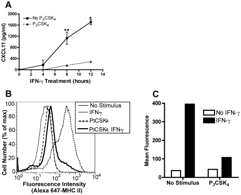
Figure 2. TLR2-mediated inhibition of IFN-γ induction of CXCL11 and CIITA decreases expression of protein products.
A. BALB/c BMDM were treated with 10 ng/ml Pam3CSK4 for 8 h followed by 20 ng/ml IFN-γ for 4, 8, and 12 h. Culture supernatants were collected and assayed for CXCL11 protein by ELISA. *, p<0.01, **, p<0.05 comparing IFN-γ alone with Pam3CSK4 and IFN-γ treated samples (as determined by two-tailed t-test). B and C. BMDM were treated with 10 ng/ml Pam3CSK4 for 12–15 h prior to stimulation with 20 ng/ml IFN-γ for 24 h. Cells were stained with Alexa 647-conjugated anti-mouse I-A/I-E and analyzed by flow cytometry. Data shown are fluorescence intensity vs. cell number (B) and mean I-A/I-E fluorescence (C). Results are expressed as means±SEM from two independent experiments (A) and are representative of at least five independent experiments (B and C).

