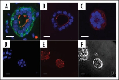
Figure 2.
Formation of acini in different matrices. (A–C) Mammary epithelial cells (EpH4) adopt the correct polarity in Matrigel™ but not in collagen gel preparations. (A) Tissue section from virgin mammary gland showing bi-layered epithelium. (B) EpH4 cells in Matrigel. (C) EpH4 cells in Collagen gel. Antibody staining: Green, cytokeratin14, a marker of myoepithelial cells; red, aquaporin 5, a marker of polarized cells that is expressed on the luminal face; blue, DAPI (nucleus—DNA). (D–F) KIM-2 cells form acini when cultured in Matrigel. (D) Nuclear staining (DAPI); (E) E-Cadherin (red) showing cell adhesions. (F) phase contrast. Scale bars = 10 µm.

