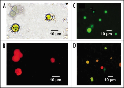
Figure 2.
(A) Recently isolated Nostoc cells from Peltigera canina thallus without any treatment, observed at light microscope. (B) The same untreated Nostoc cells observed under fluorescence microscope. (C) Isolated Nostoc cells incubated for 2 h in the dark with 20 g of FITC-arginase secreted from P. canina thalli and observed under fluorescence microscope. (D) Isolated Nostoc cells incubated for 2 h in the dark with 20 g of FITC-arginase secreted from P. canina thalli, for 1 h on 100 mM α-D-galactose in 0.1 M phosphate buffer, pH 7.4, and observed under fluorescence microscope.

