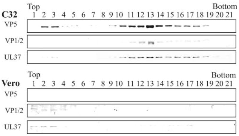
FIG. 6.

Rate-zonal centrifugation of capsids isolated from cells infected with the VP23-negative mutant (K23Z) and analysis for the presence of tegument proteins VP1/2 and UL37. Vero (non-complementing cell line) and C32 (complementing cell line expressing VP23) cells were infected with the VP23-negative mutant virus, K23Z, at a MOI of 10 pfu/cell. At 15 h after infection the capsids from the nuclear fraction were obtained, separated by rate-zonal centrifugation, analyzed by SDS-PAGE followed by Western blot analysis as described in the legend to Fig. 2. The same blot was probed sequentially for the presence of VP5 and tegument proteins VP1/2 and UL37.
