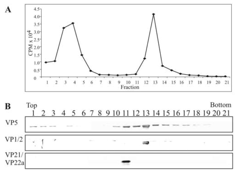
FIG. 7.

Rate-zonal centrifugation of 3H-thymidine-labeled capsids and analysis for the presence of the tegument protein VP1/2. Capsids were obtained from the nuclear fraction of HSV-1-infected Vero cells that were labeled with 10 μCi per ml of 3H-thymidine from 3 to 15 h after infection. The capsids were separated by rate-zonal centrifugation as described in the legend to Fig. 2. Prior to TCA precipitation of the proteins within each of the fractions, 50 μl was removed and 3H-thymidine cpm in each fraction were determined by liquid scintillation analysis (panel A). Following TCA precipitation, the same fractions were resolved by SDS-PAGE followed by Western blot analysis (panel B). The same blot was sequentially probed with antibodies to the tegument protein VP1/2 and capsid proteins VP5 and VP21/VP22a (scaffold proteins) as described in the legend to Fig. 2.
