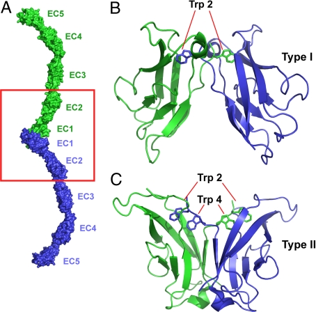
Fig. 1.
Structure of cadherin molecules. (A) The structure of the adhesive dimeric complex formed by ectodomains of type I C-cadherin (7) is shown in molecular surface representation. Note that the adhesive interface is encompassed entirely within the EC1 domain. The red boxed region includes the interacting EC1–2 regions corresponding to the cadherin constructs used in this study. (B) Expanded ribbon diagram view of the strand-exchanged adhesive interface between the N-terminal EC1 domains of the type I E-cadherin (9). The side chains of the Trp anchor residues are shown. (C) A ribbon diagram view of the strand-exchanged adhesive interface between the N-terminal EC1 domains of the type II cadherin 8 (15). The side chains of the Trp-2 and Trp-4 anchor residues are indicated.

